
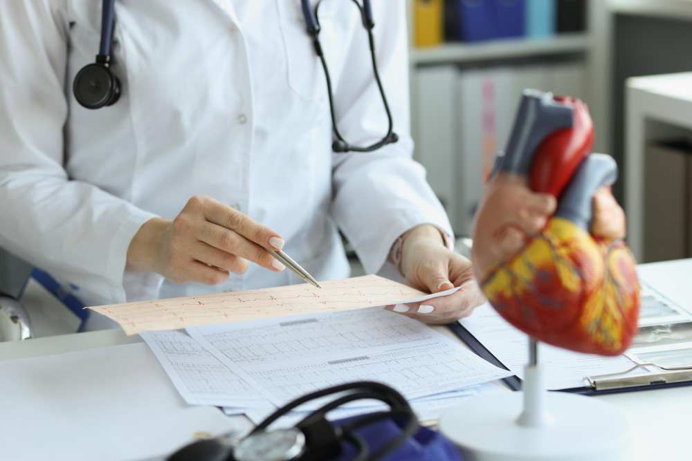
Evaluation of each heart problem starts with a good doctor-patient relationship. There should be a two-way conversation. Listening to the whole story is always the first and most important step. The symptoms, past medical history, family history, drugs used, lifestyle, diet and habits (smoking, drinking alcohol, illicit drugs), occupation and hobbies all matter while evaluating a patient. Then, we proceed to a detailed systemic and cardiovascular physical examination and come up with a differential diagnosis.
To support the most probable diagnosis and to exclude the rest, we have to do certain medical tests. Usually, the heart tests are chosen according to the pretest probability of the patient to certain cardiac diseases (e.g: if a patient is at intermediate risk of having a future heart attack, it is logical to order a treadmill exercise test (TMT). Still, for an 18-year-old healthy female whose pretest probability of having significant coronary artery disease is nearly zero, the chance of having false negative results will be very high) and patient characteristics(e.g: if a patient is obese or cannot walk, you cannot order a TMT).
Heart Tests Recommended by Cardiologist to Diagnose Heart Problems
Cardiologists may recommend the following tests to assess heart health and diagnose potential problems:
1. Blood tests
Blood tests help diagnose heart attack, high cholesterol, thyroid hormone disorders, fatty liver, and microalbuminuria in patients with hypertension and diabetes. They can also identify certain risk markers of coronary artery diseases like homocysteine and high-sensitive CRP. Patients with heart failure and under high-dose diuretic therapy should have their blood tests done periodically to detect any electrolyte abnormalities.
2. Chest X-ray
Chest X-rays provide insights into the heart’s shape, size, position, as well as associated abnormalities in the lung fields and vascular structures. Its uses in cardiology are as follows:
- Increased cardiothoracic ratio (CTR) is a sign of heart enlargement. It is used to diagnose heart failure, cardiomyopathy( heart muscle disease), and valvular heart diseases.
- Venous congestion in the lungs can indicate left-sided heart failure and dilated lung arteries may be seen in pulmonary arterial hypertension.
- Pleural effusion( fluid collection in lungs), lung edema or vascular redistribution in the lungs can be observed, which are indicative of heart failure or other conditions leading to fluid overload.
- Chest X-rays are crucial for evaluating the position and integrity of cardiac devices such as pacemakers, defibrillators(ICD) and metallic heart valves.
- Although echocardiography is the main tool for the diagnosis of congenital heart diseases, chest X-rays can still provide useful information and clues. ( ex. Boot-shaped heart in tetralogy of Fallot).
- Helps exclude other causes of chest pain and shortness of breath, such as pneumonia, lung tumours, and pulmonary embolism.
3. Holter monitoring
Holter is a device that takes ECG for a long period and stores the data. A normal ECG can miss arrhythmias that are not persistent but intermittent. Patients with suspected intermittent arrhythmias can be monitored with a 24, 48, or 72-hour Rhythm Holter. The data obtained during these hours are stored and transferred to a computer, which a cardiologist then reads. Neurologists also frequently use Rhythm Holter to detect paroxysmal atrial fibrillation( an irregular rhythm in elderly patients associated with stroke) in patients with stroke.
4. Electrocardiogram (ECG or EKG)
ECG records the electrical activity of the heart from electrodes (10 electrodes – 4 extremities and 6 chests) attached to the patient’s body. ECG provides information about heart rate, basal heart rhythm, and heart axis, helps to diagnose ST-segment elevation heart attacks before blood test results, diagnoses arrhythmias, and also gives information about other cardiovascular diseases like hypertension, past heart attacks, coronary artery disease, heart valve disease, pulmonary hypertension, pulmonary embolism. It also protects us from conducting unnecessary tests and saves time and money.
5. Tension Holter
Some patients have high blood pressure at the hospital and normal at home. Some may have only occasional high blood pressure, while others may have high blood pressure only at night. Tension Holter helps diagnose real hypertension, exclude pseudo hypertension and sometimes diagnose the type and cause of hypertension (e.g. white coat hypertension- patient only has high blood pressure in front of the doctor but normal BP in day to day life, stress-induced hypertension, sleep apnea-induced hypertension)
6. Exercise Stress Test (Treadmill Stress Test- TMT)
In TMT a patient runs on a treadmill with speed and inclination increasing every three minutes. We increase the oxygen demand of the heart muscle and check whether the coronary artery blood supply is enough. When there is a supply-demand mismatch, subendocardial ischemia occurs, which is presented either by chest pain or ST segment depression in an ECG.
From the history and physical examination, we classify a patient in a risk group for coronary artery disease. The TMT has a specificity and sensitivity of 70-80%. That’s why this test is suitable for patients who are at intermediate risk for coronary artery disease. Exercise stress is also used in various other conditions like preoperative risk assessment, assessment of severity of heart valve problems, and functional class assessment after a massive heart attack.
7. Transthoracic Echocardiography (TTE)
TTE is a non-invasive procedure that involves placing an ultrasound transducer on the chest or abdomen. Ultrasonographic waves echo off the heart structures and return to the transducer, which then converts them into real-time images, allowing clinicians to view the heart’s chambers, valves, and surrounding areas.
TTE is one of the most used diagnostic tools in cardiology and helps to determine cardiac function (contractile power, ejection fraction), diagnose valvular heart diseases, congenital heart diseases, shunts (intracardiac and between major vessels), anatomy and dimension of major vessels (aorta and pulmonary artery), aortic dissection flap, pulmonary embolism (right heart dilation), pulmonary hypertension and its causes, previous heart attack, coronary artery disease (contractility abnormality in certain wall segment), and complications after heart attack (valvular insufficiencies, chordal rupture, pericardial effusion, etc.).
8. Transesophageal Echocardiography (TEE)
Unlike a traditional TTE where the transducer is placed on the chest, TEE involves inserting a transducer on the tip of a flexible tube through the mouth and into the esophagus. This positioning allows for clearer images of the heart because the esophagus is directly behind the heart, bypassing barriers like skin, muscle, and bone which can obstruct or distort the ultrasound waves in TTE.
TEE is used to investigate the presence of thrombus or mass in a stroke patient, vegetation and valvular complications in infective endocarditis, calculate the function of the prosthetic valves, diagnose aortic dissection, identify and evaluate intracardiac shunts( ASD, VSD, PFO), to determine the efficacy of cardiovascular surgery(e.g., mitral valve repair).
It is also used to diagnose congenital heart disease and as a guide in the treatment of structural heart disease like PFO Closure, ASD Closure, VSD Closure, Appendix Closure, Paravalvular Leak Closure, Mitraclip, Tricuspid clip etc.
9. Coronary Angiogram (Cardiac Catheterization)
With the help of radiopaque contrast injection to the targeted vessel and visualization of the vessels using an x-ray, the angiography is performed. This helps to detect any problems inside the lumen of the vessels.
Coronary angiography is the gold standard tool to diagnose coronary artery disease. Moreover, it is used to visualize carotid arteries, intracranial arteries, the aorta and its branches, upper and lower extremity arteries, renal arteries, pulmonary arteries etc.
10. Nuclear Cardiac Stress Test (Myocardial Perfusion Imaging – MPI)
In MPI heart muscle oxygen demand is increased by exercise or drug injection causing increased heart rate and perfusion of the heart muscle is calculated. If the perfusion defect is found in >10% of heart muscle territory, the patient is considered at high risk for future cardiac adverse events and should undergo coronary angiography. Besides, MPI is used to detect viable tissue in patients who have had heart attacks. This helps us to decide whether opening the blocked vessel is beneficial for the patient or not.
11. Magnetic Resonance Imaging (MRI)
Cardiac MRI is used to diagnose certain diseases that affect the muscle of the heart (myocarditis, cardiomyopathies, infiltrative diseases), and diagnose the presence of scar tissue and hence the risk of death in certain high-risk patients with episodes of fatal arrhythmias( ventricular tachycardia, ventricular fibrillation etc.). Cardiac MRI can provide static, dynamic, and functional information about the heart and valves. Although it is becoming popular day by day, its use in daily practice and unstable patients is not feasible because of the time consumed and the expertise needed both to perform and interpret the test.
12. Coronary Computed Tomography Angiogram (CCTA)
CT angiography is widely used to see coronary anatomy. In patients with severe coronary calcification, multiple stents and unstable heart rate( atrial fibrillation), the results might not be accurate. Therefore, the test should be ordered according to the patient’s age, patients clinical condition and the pretest probability of coronary artery disease.
13. Coronary Artery Calcium (CAC) Test
CAC is a non-invasive imaging test that uses computed tomography (CT) to measure the amount of calcified plaque in the coronary arteries. The primary aim is to evaluate the risk of developing coronary artery disease (CAD) by quantifying the amount of calcium deposits in the arteries that supply blood to the heart muscle. Higher amounts of calcium suggest more severe disease and increase the risk of heart attacks or other cardiovascular events.
14. Tilt-Table Test
Patients with recurrent episodes of syncope (fainting) but without any structural heart disease and obvious cause can be selected for a tilt table test to identify the cause and classify the type of syncope.
15. Nuclear Imaging Tests
This test is already explained above.
Conclusion
While developing the medical tests, they are always used to detect disease in those people who are at risk of having it. Let me give you an example: If you use an exercise test for a 50-year-old male, smoker with and history of heart attacks in the family then the positive test means he may have a problem with his heart. If you do the same test on a 20-year-old boy, then the positive test actually means the disease is less than 5%. So the test results should always be correlated with patient symptoms and story. Ensure your heart health with the right tests and expert guidance. Schedule your Online consultation today with the best heart doctor to discuss the best diagnostic options for you.

Related Blog Post: Read More About Heart Attack
- Heart Attack Early Warning
- Heart Healthy Diet Plan After a Heart Attack
- What to do When Heart Attack Occurs
- How to Avoid Heart Attacks with a Healthy Lifestyle
FAQs on Medical Tests to Diagnose Heart Problems
1. What is the most effective test for heart disease?
There is not any single test that is most effective for all heart disease. As discussed earlier, tests should always be ordered according to patients’ symptoms, story and cardiovascular risk profile. In cardiology, echocardiography is the most extensively used test.
2. Is ECG enough to detect heart problems?
ECG provides valuable information about heart rate, heart rhythm, heart axis and helps to diagnose heart attacks and arrhythmias. It also gives information about other cardiovascular diseases like hypertension, past heart attacks, coronary artery disease, heart valve disease, pulmonary hypertension, pulmonary embolism, and ST-segment elevation heart attack. However, ECG alone is not enough to detect all heart problems. Usually, one or more additional tests are needed to diagnose heart diseases.
3. Which test shows your heart is healthy?
Overall heart health cannot be identified by a single test. If a symptom-free patient has normal ECG, normal blood tests, normal TMT with good functional capacity and normal echocardiography, then we can say their heart is healthy.
4. Can an MRI show blocked arteries?
MRI is not the diagnostic tool to detect blocked coronary arteries. Although specific MRI techniques could identify coronary artery blockages with an accuracy of 88% when compared to CT coronary angiography, it takes much more time and needs expertise and patient stability. So it is not used for this purpose in clinical practice.
5. What is the cost of a cardiac MRI?
The cost of cardiac MRI depends upon countries, machines used and experts interpreting the result. The cost is the highest compared to all the above-mentioned cardiac tests.
6. Which is better, an ECG or CT scan?
ECG is better at evaluating rhythm disorders and immediate diagnosis of an ST-segment elevation heart attack whereas a CT scan is better at evaluating heart anatomy, coronary artery anatomy, stenosis and extension of coronary diseases.